The use of X-rays in the dental clinics is fortunately becoming more and more widespread. This technique, which has been helping to save human lives for decades, is also very useful, sometimes even essential, for the practice of dentistry. However, its use is always surrounded by controversy, due to the link between radiation and certain types of cancer.
Therefore, in Velez & Lozano we want to talk today in this blog post about X-rays, their real impact on health and the different types we use in our clinic and why:
X-RAYS AND THEIR IMPACT ON HEALTH
Radiation is the emission, propagation and transfer of energy in any medium in the form of electromagnetic waves or particles. In other words, the heat provided by the sun, which enables life on earth, is a form of radiation, as is the radiation emitted by the radio and the microwave oven we use to heat the milk for breakfast.
In fact, radiation considered to be of natural origin accounts for 64,86% of the radiation received by the Spanish population each year, according to the data handled by the United Nations Scientific Committee and offered by Spain's Nuclear Safety Council on its official website. These radiations are those coming from space (such as the example we have given about the sun), called 'cosmic radiations'; those coming from radon gas, which comes from uranium and is found naturally all over the planet; those coming from gamma rays emitted by certain materials (with which even buildings are constructed) and those emitted by certain food components, such as potassium.
Thus, although today there are mostly conspiracy movements attributing a multitude of relatively contemporary problems to radiation, there is no such thing as a 'radiation-free life'.
If we go back to the data, the average Spaniard annually receives a total of 3.7 mSv -miliSievert, a measure used to measure radiation-, while a dental X-ray emits 0.016 mSv, according to the average drawn up by the United Nations. In other words, a dental X-ray emits the 0'42% of the radiation we endure per yearThe impact on the total is therefore practically non-existent.
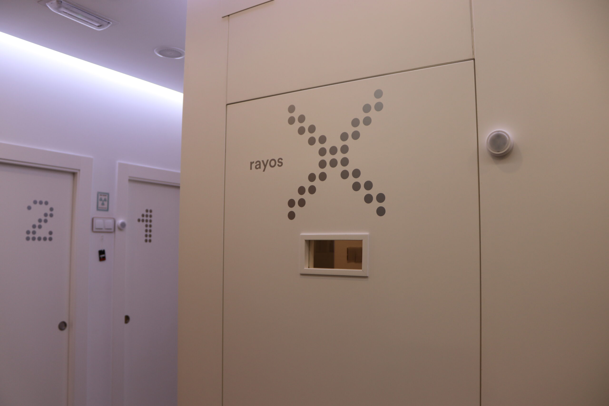
TYPE OF DENTAL X-RAYS
In our dental clinic in Murcia we use various types of X-rays, and we do so to achieve different results. Without going into too much technical detail, we will explain more or less what each one consists of:
Orthopantomography
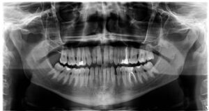
The orthopantomography is a type of X-ray that provides very sharp images of the arches of the mouth, making it possible to detect possible anatomical, structural, bone or dental damage.
Most patients often confuse it with CT or CBCT, as they are performed with the same machine and have a similar procedure for them, which requires them to bite down on a tongue and rest their jaw on a support while holding two handles with their hands.
Teleradiography
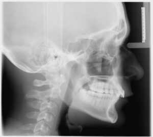
This test shows the patient's profile and is performed almost exclusively for the practice of orthodonticsIt is also a fundamental test for orthognathic surgery, at least in our clinic, but it is also a fundamental test for orthognathic surgery, for example.
Cone Beam Computed Tomography (CBCT)
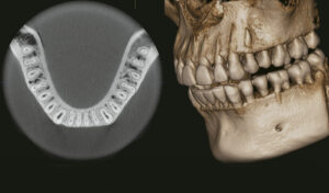
Although many patients identify it as a CT scan, the test performed at Vélez & Lozano is actually a CBCT. The fundamental difference is that X-ray computed tomography is emitted in the shape of a cone, whereas CT is cylindrical. This modern technology makes it possible to form a 3-dimensional image of teeth, soft tissues, bones and nerves in a single scan.
It is often used primarily and fundamentally in surgeries, such as implant placement.
Bite Alert
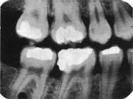
This is one of the most versatile and common tests performed in dental clinics. In this test, the patient holds the X-ray plates inside his or her mouth and the X-rays are taken from the outside using an X-ray machine.
It is very useful for the detection of interdental caries and assess the condition of the crown or periodontal tissue, among others, hence they are frequently performed.
Periapical radiography
This technique is performed in much the same way as the flap technique, but in this case the radiographic plates are positioned so that the apexes, the tip of the roots and the area around them can be seen after development.
What is sought is greater definition of certain details that may not have been clear after an ortho.
For more information, you can always visit our dental clinic in Murcia, where we will be happy to help you. We also attend by phone on 968 28 46 28 or through our social networks.
Author: Vélez&Lozano