As promised in my last article about magnifying loupes, the use of the microscope in dental clinics such as Vélez y Lozano is a topic that could be the subject of a new article in itself. This one in particular.
Let's start at the beginning, with a bit of history, which will allow us to increase our knowledge of the subject in the right way.
History and application of the optical microscope in dentistry
Although what is considered to be the first microscope in history was built by two Dutch opticians, Zacharias and Hans Janseen, in 1590, it was not until 1953 that the binocular operating microscope as we know it was marketed to the public, and was also offered for sale in the Netherlands.
The introduction of the optical microscope in medicine occurred a few years later, in 1957, and ear specialists were the first specialists who began to use it in their practices, although it did not take long for it to spread to ophthalmology, neurosurgery, plastic surgery and microsurgery. At the end of the 1970s, the first publications of scientific literature were found in which the benefits that the use of an apparatus such as the microscope could represent for dentistry, specifically for the performance of surgical endodontics.
Thus, in 1978 Apotheker and Jako had the idea to introduce this new device into the dental field and collaborated in the development of the first dental operating microscope, which was marketed in 1981 by the Dentiscope company, but its use remained rather restricted for decades.
It was not until 1995 that the American Association of Endodontics (AAE) recommended that microscopy training be included in the Advanced Endodontic Education Programmes. In January 1997, training in the use of the microscope was declared mandatory for the specialty.
Why is the use of an optical microscope in dentistry recommended?
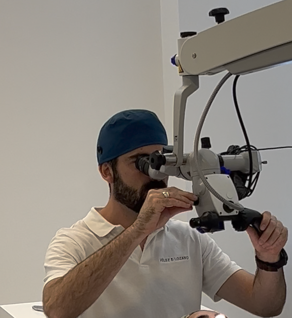
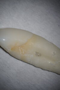
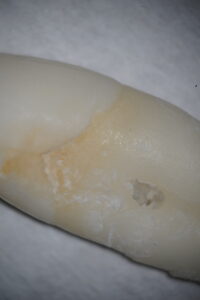
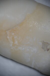
In certain cases, due to their level of complexity and the need to be aware of details that the human eye is not able to capture, optical microscopes can be of great help. In addition to allowing us a very good ergonomic posture, they allow us to have magnifications that are best described with the help of images.
Of course, the depth of field and light input is also considerably reduced, so the microscopes have a powerful light that allows you to appreciate the details at the highest magnifications. In our dental clinic in Murcia we have one of the Zeiss brand, which guarantees the highest levels of quality.
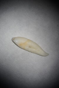
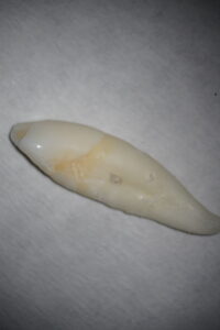
Let's look at increases with graphic examples:
It should be noted that the focal length of the microscope is fixed. It also has an output for attaching a camera to document the treatment. This is not only used for didactic purposes in internal clinical sessions, many colleagues use it for training courses or to collect information for scientific dissemination. In addition, it can be shown to the patient to explain something about the treatment or it can be projected on television so that the patient's companion can see the procedure being performed live.
The use of the microscope in dental clinics
Although we know the theory, in practice in Spain, the use of microscopes in dental clinics is not really widespread. In fact, according to a study published in the Journal of the Ilustre Consejo General de Colegios de Odontologos y Estomatologos de España74.66% of the collegiate respondents, from different specialities, did not use a microscope.
A very high figure that is undoubtedly due to the lack of availability in most dental clinics in Spain, which in turn is due to the high investment required.
We are aware that this type of investment in technology that we do make at Vélez y Lozano is what, among other factors, makes us stand out as a leading dental clinic in Murcia.